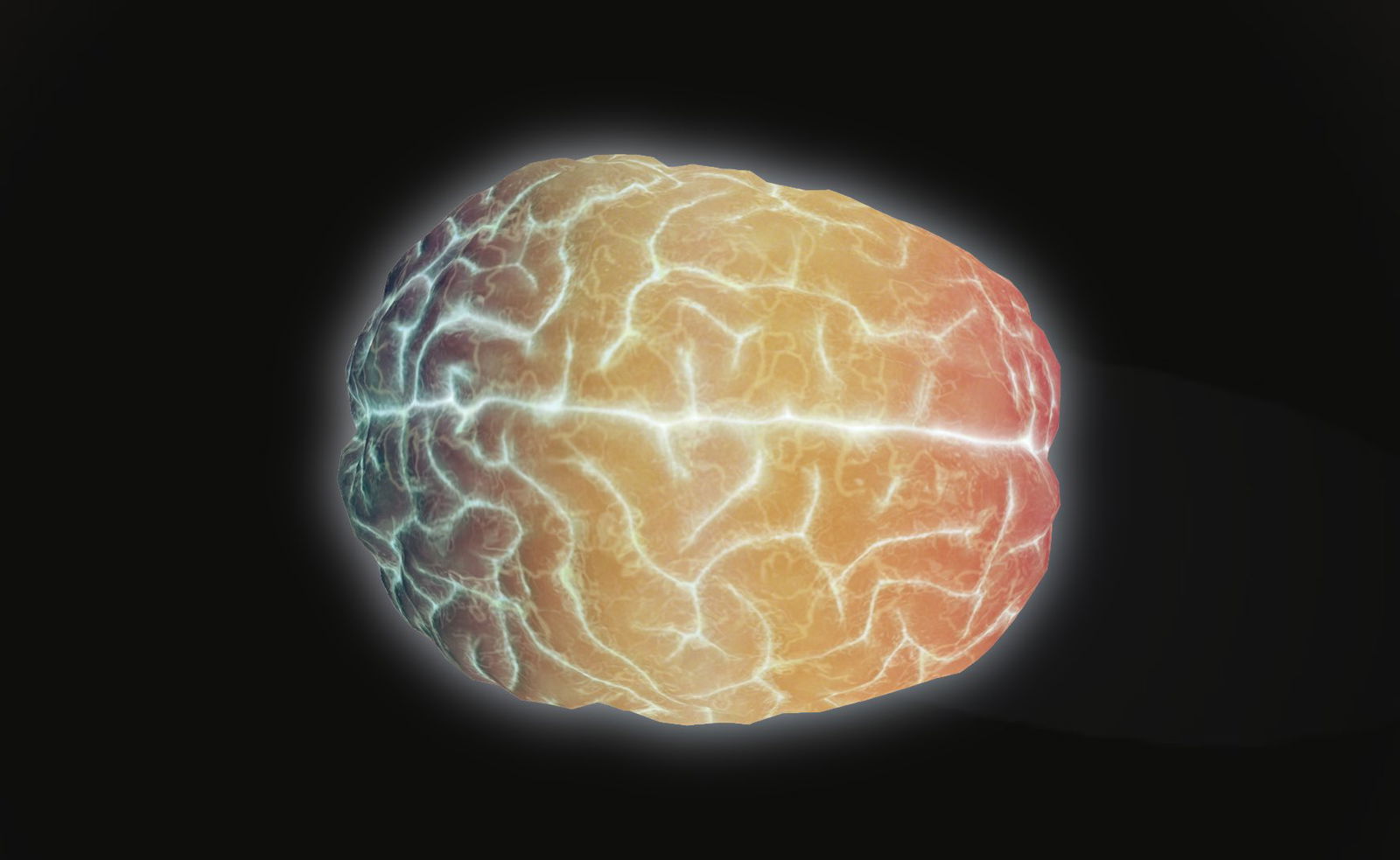
Physicists have demonstrated the first successful detection of light transmitted through an entire adult human skull, overcoming attenuation levels previously considered impossible.
Published in Neurophotonics, the study reveals how carefully engineered laser systems and single-photon detection methods enabled researchers to track photons traveling 15.5 cm across a human head—a feat with profound implications for non-invasive brain scan technology.
The team used a special pulsed laser and a time-correlated single-photon counting system to detect photons surviving a journey through skin, skull, cerebrospinal fluid, and brain matter. Despite attenuation equivalent to just one photon surviving per quintillion emitted, the system recorded approximately one photon per second after 30 minutes of data collection.
So what exactly does this all look like?
In a setup that could double as the world’s most precise game of laser tag, researchers fired pulses of light at one side of a participant’s head while an ultra-sensitive detector waited opposite, ready to catch photons that survived the journey. Blocking all other light in the room, the researchers then waited to see if the detector began to pick up any photons.
Previous computer simulations indicated that light would take certain routes through the brain in order to make it out the other side, and the experimental results came incredibly close. While photons did manage to make it through the dense and complex brain material, and exit the other side, they generally preferred to travel via the cerebrospinal fluid (which is the clear protective fluid that surrounds the brain and spinal cord).


However, significant barriers remain.
Successful detection occurred only in a fair-skinned, hairless participant among eight test subjects, highlighting biological variability in optical properties. The 30-minute acquisition time and reliance on 1.2 W lasers, which are near the safety limits for skin exposure, also pose practical challenges. Suffice it to say, while blasting someone’s head with a laser worked, it needs some fine tuning to become viable for medical bedside use.
That being said, this research establishes a framework for developing optical systems that combine the portability of EEG with the depth resolution of fMRI, which could be a significant boon for global healthcare accessibility. As computational methods and detector technologies advance, the dream of affordable, non-invasive deep-brain imaging appears increasingly within reach.
MJ Banias covers security and technology with The Debrief. You can email him at mj@thedebrief.org or follow him on Twitter @mjbanias.