
Researchers at Harvard and Google have produced the most detailed map of the human brain ever rendered, revealing the complicated neural interactions occurring within a tiny cubic millimeter sample of brain tissue.
The collaborative effort showcases the power of current electron microscopy paired with the ever-growing capabilities of artificial intelligence, and offers unique insights into neural connections in vivid imagery made available online by the researchers.
Within a single cubic millimeter of brain tissue are close to 57,000 cells, along with hundreds of synapses and blood vessels that, when reproduced in the Harvard and Google collaboration’s reconstructions, resulted in close to 1,400 terabytes of data.
The groundbreaking effort was led by Jeff Lichtman, Harvard’s Jeremy R. Knowles Professor of Molecular and Cellular Biology and newly appointed dean of science, offering an unprecedented glimpse at the intricacies of the human brain and its inner workings.
A decade in the making, Lichtman and the Google team’s research combines electron microscopy with Google’s advanced AI in a meticulous reconstruction of the complexities of the brain, as outlined in a recent scientific study by Lichtman, along with former Harvard postdoctoral researcher Alexander Shapson-Coe, Michał Januszewski of Google Research, and Harvard postdoctoral researcher Daniel Berger as co-first authors.
Now, the newly unveiled brain map, which the researchers call a “connectome,” has revealed what Lichtman recently characterized as “an alien world inside your own head.”
In the newly published study, the authors say the functions occurring within our brains represent the key difference that separates us from other life on Earth. Despite this recognition, we currently possess a less-than-complete understanding of the intricate synaptic circuitry underlying these functions.
Enter Google Research’s Connectomics team, which, over the last decade, has worked toward using machine learning to construct a clearer picture of the human brain at previously unmatched scales. The result of this work allows researchers to produce visualizations of extremely large volumes of neural circuits at high resolutions, which could lead to entirely new discoveries about the way our brains function.
A limiting factor in this research involves the problems with obtaining high-quality samples of human brain tissue. Unlike other organ biopsies, brain biopsies rarely occur except when used to extract neoplastic masses, which aren’t ideal for creating visualizations of normal brain structure. In past attempts, brain organoids composed of human cells have been used, but since they lack cortical layers, they were unable to faithfully reproduce all of the features of real human brain tissue.
In their recent paper, the study’s authors proposed a new strategy involving the use of human brain tissue extracted during neurosurgical procedures, which they said “could be leveraged to study normal—and ultimately disordered—human neural circuits.”
Lichtman obtained such a sample a decade ago. Smaller than a single grain of rice, the sample had been retrieved from the brain of a patient with severe epilepsy as part of a standard procedure where a small portion is removed to help reduce seizures and then analyzed to determine whether it is normal. Apart from the patient’s age and gender, Lichtman knew nothing else about the sample’s origins.
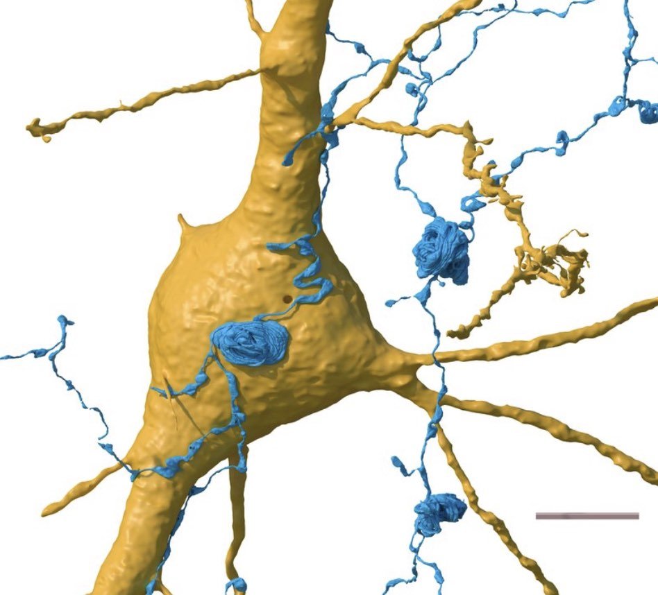
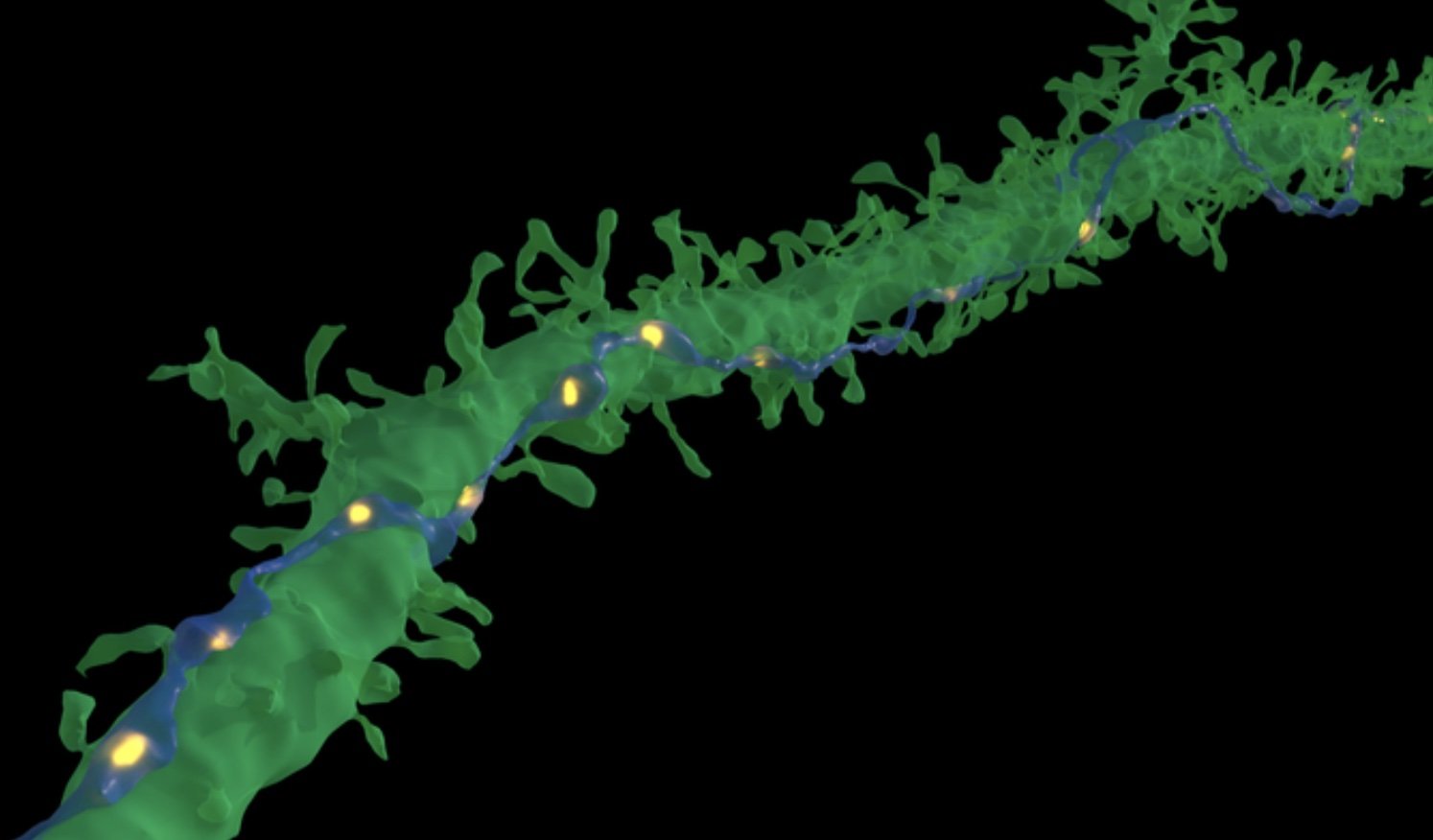
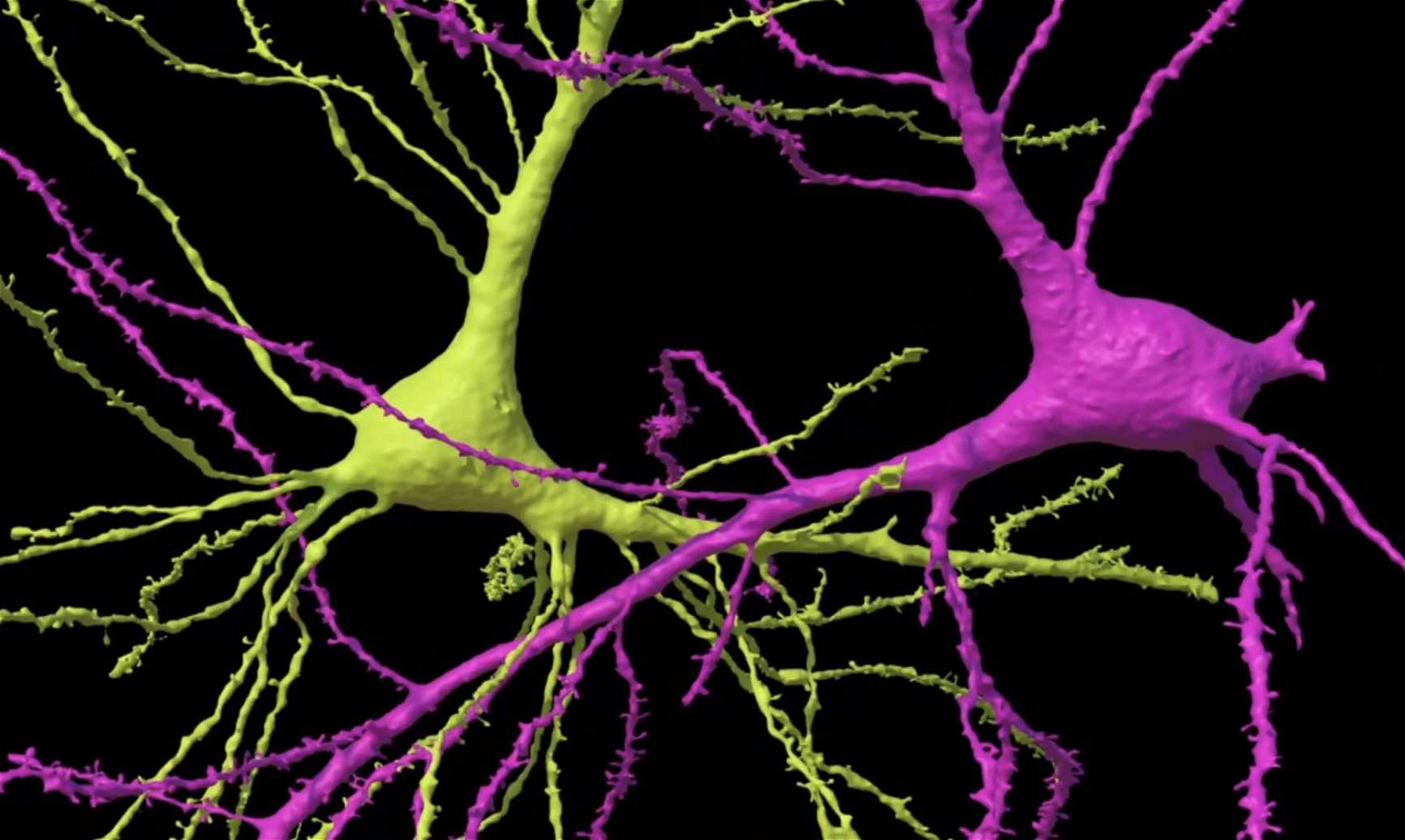
In their research, the team used electron microscopy to produce a massive set of very detailed images using this small piece of temporal cortex, which was only about 1 cubic millimeter in size. Then they analyzed the data with Google’s powerful AI, reconstructing thousands of neurons, over a hundred million synaptic connections, and other brain components like glial cells, blood vessels, and myelin.

The tremendous resulting trove of data, which revealed a previously unrecognized class of neurons hidden within deep layers of the tissue and “very powerful and rare multisynaptic connections between neurons throughout the sample,” was made available online, along with tools to aid other researchers in analyzing it.
“This work provides evidence of the feasibility of human connectomic approaches to visualize and ultimately gain insight into the physical underpinnings of normal and disordered human brain function,” the researchers write in their new study. “It is hoped that this endeavor will be aided by providing free access to all of the data and relevant tools.”
“Given the enormous investment put into this project, it was important to present the results in a way that anybody else can now go and benefit from them,” said Google Research collaborator Viren Jain of the team’s findings.
Supported by the National Institutes of Health BRAIN Initiative, this collaboration’s ultimate goal is to create a high-resolution map of a whole mouse brain’s neural wiring. This ambitious project will require approximately 1,000 times the data produced from the current 1-cubic-millimeter fragment of human cortex used by Lichtman and his collaborators in the recent research.
The team’s landmark paper, “A petavoxel fragment of human cerebral cortex reconstructed at nanoscale resolution,” was published in the journal Science on May 10, 2024.
Micah Hanks is the Editor-in-Chief and Co-Founder of The Debrief. He can be reached by email at micah@thedebrief.org. Follow his work at micahhanks.com and on X: @MicahHanks.