In a groundbreaking new study, researchers have achieved something straight out of science fiction by turning living skin transparent.
Using an innovative approach involving a common food dye, scientists could temporarily render the skulls and abdomens of live mice transparent, allowing them to peer into the inner workings of their bodies without invasive surgery.
This development, published in the latest issue of Science, could have far-reaching implications for biomedical research and the study of complex biological systems.
“For those who understand the fundamental physics behind this, it makes sense; but if you aren’t familiar with it, it looks like a magic trick,” Dr. Zihao Ou, an assistant professor of physics at The University of Texas at Dallas and lead author of the study said in a statement.
Optical imaging is a critical tool in modern biology and medicine, enabling researchers to study living organisms in real-time. However, the natural opacity of biological tissues poses a significant challenge to scientists seeking to observe structures deep within the body.
Traditional optical imaging methods are often limited by light scattering and absorption, which restricts the depth and clarity of the images that can be obtained.
The new study, led by Dr. Ou, aimed to overcome this hurdle by developing a novel technique for making biological tissues transparent.
“The desire to see inside biological tissue and uncover the fundamental processes of life has spurred extensive research into deep-tissue optical imaging methods, such as two-photon microscopy, near-infrared-II fluorescence imaging, and optical tissue clearing,” researchers wrote. “However, these methods either lack sufficient penetration depth and resolution or are unsuitable for living animals.”
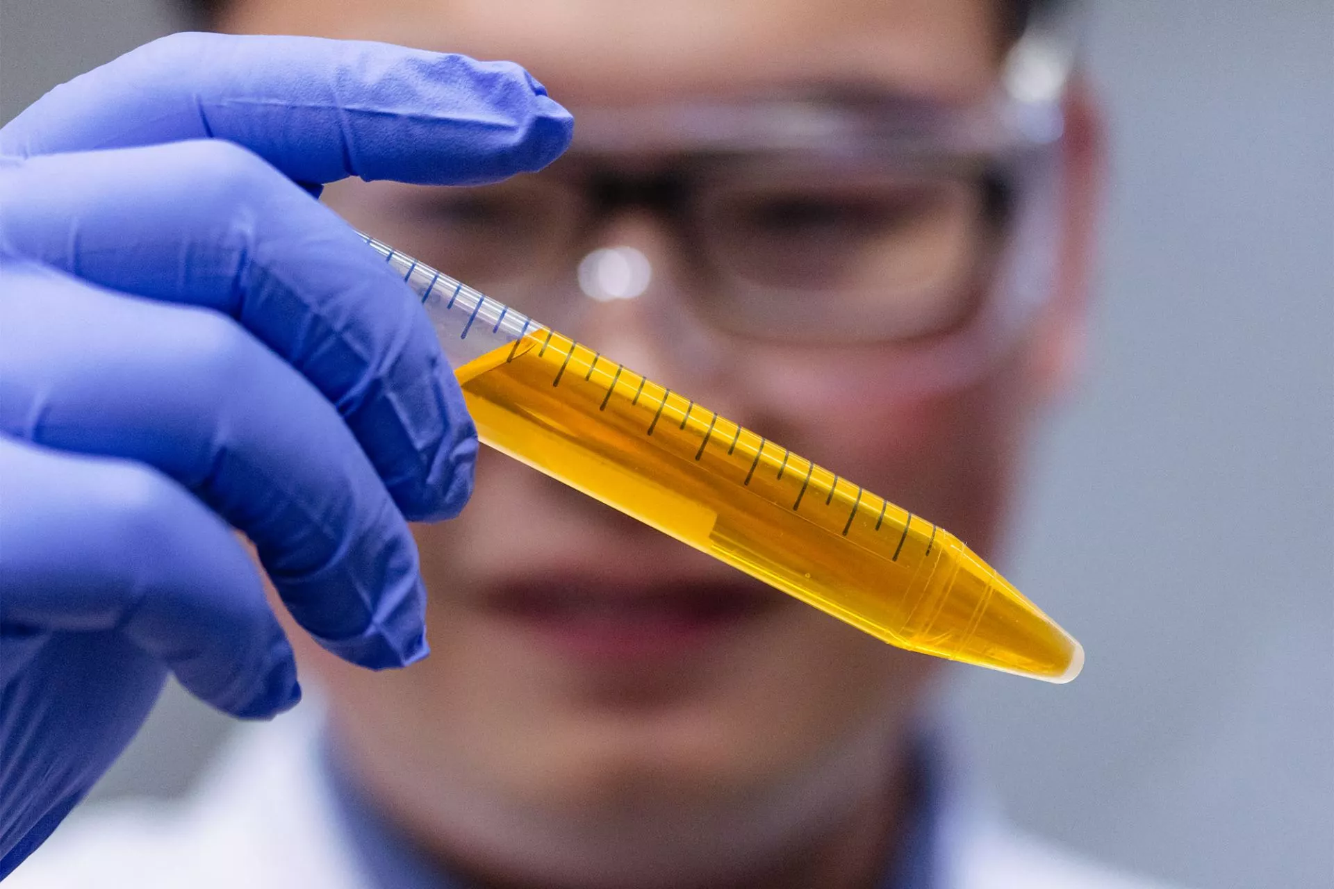
Researchers devised a surprisingly elegant solution by using a common yellow food dye known as tartrazine.
Approved by the U.S. Food and Drug Administration, tartrazine is widely used in everyday products, from candies to soft drinks. However, this study used it for a very different purpose.
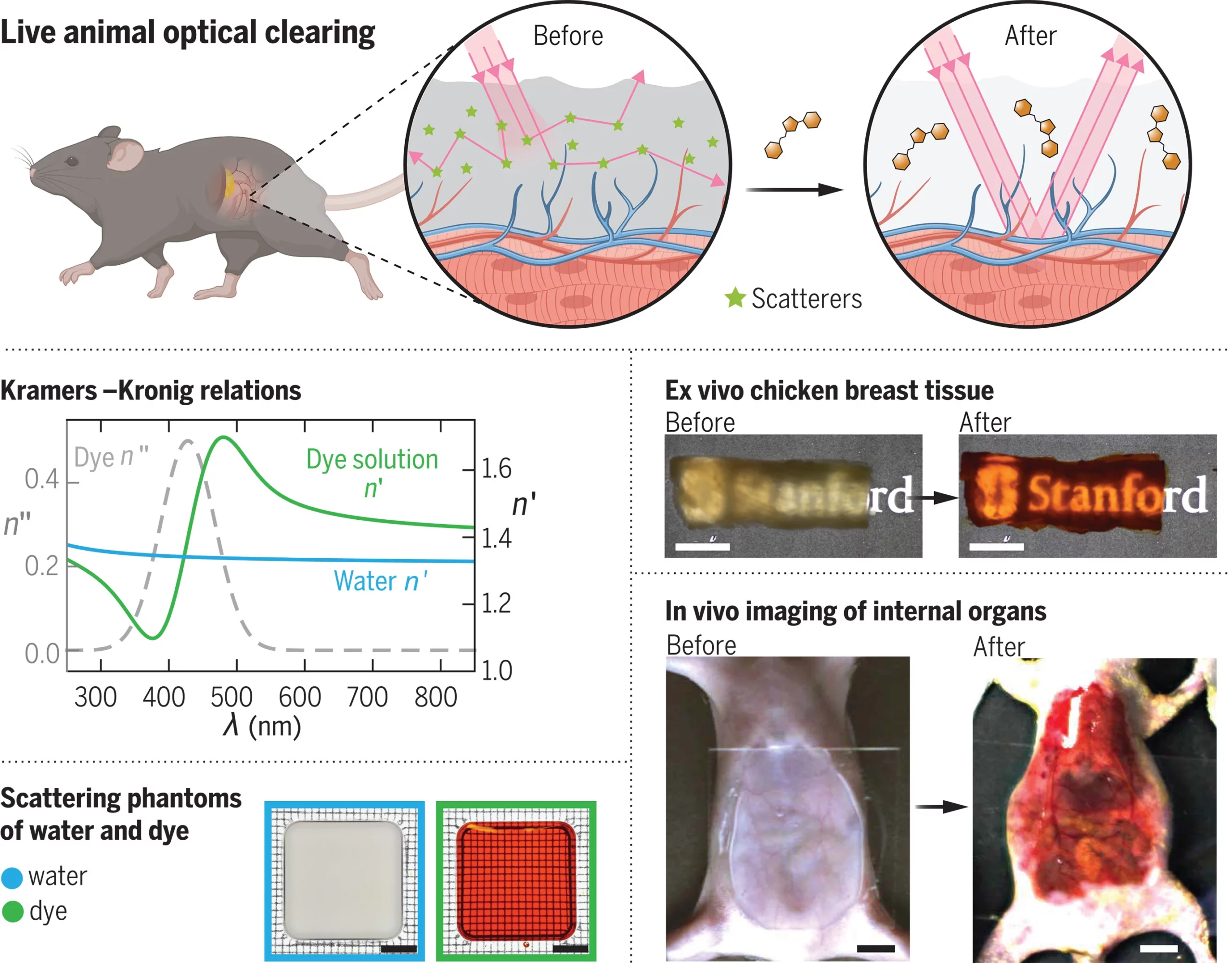
The team found that when dissolved in water, this dye could selectively absorb light in the blue region of the visible spectrum. This absorption effectively alters the refractive index of the aqueous medium, increasing transparency in the red part of the spectrum.
“We combined the yellow dye, which is a molecule that absorbs most light, especially blue and ultraviolet light, with skin, which is a scattering medium,” Dr. Ou explained. “Individually, these two things block most light from getting through them. But when we put them together, we were able to achieve transparency of the mouse skin.”
The study’s theoretical framework relies on the Lorentz oscillator model, a well-known concept in physics that describes how light interacts with matter.
By applying this model, the researchers predicted that certain dye molecules could enhance transparency in biological tissues. The experimental results confirmed these predictions, demonstrating that tartrazine could temporarily make live mice’s skin, muscles, and connective tissues transparent.
In a series of experiments, the team applied tartrazine to the abdomens and skulls of live mice, successfully rendering these areas transparent. This breakthrough allowed them to observe fluorescently labeled cells and structures deep within the tissues in real-time.
One of the most exciting applications was the visualization of the enteric neurons in the guts of mice. Researchers could see the neurons’ movements as they mirrored gut motility, generating real-time maps of gastrointestinal activity.
The study also demonstrated the versatility of this technique by applying the dye solution to other parts of the mouse body. For instance, researchers applied the solution to the scalp to visualize cerebral blood vessels and to the hindlimbs for high-resolution imaging of muscle fibers.
In all cases, the dye made it possible to see through the typically opaque tissues with unprecedented clarity and detail.
The process is also easily reversible by washing off any remaining dye, while the dye that has diffused into the skin is metabolized and naturally excreted through urine.
“It takes a few minutes for the transparency to appear,” Dr. Ou explained. “It’s similar to the way a facial cream or mask works: The time needed depends on how fast the molecules diffuse into the skin.”
(Image Source: Dr. Zihao Ou. et al.)
The implications of this research are vast. Making tissues transparent could revolutionize many areas of biomedical research. It could enable scientists to study complex tissue structures, such as the brain and the vascular system, in ways that have never been possible before.
Moreover, using biocompatible dye to make living skin transparent makes the optical imaging process safe, affordable, and efficient.
“In human medicine, we currently have ultrasound to look deeper inside the living body,” Dr. Ou remarked. “Many medical diagnosis platforms are very expensive and inaccessible to a broad audience, but platforms based on our tech should not be.”
The technique could be adapted for use in other animals, potentially including humans, though much more research is needed to assess its safety and effectiveness in larger organisms.
However, the researchers also note some limitations of their approach. While the dye improves transparency by reducing light scattering, it does not eliminate it entirely. The technique’s effectiveness also depends on the diffusion of the dye within the tissue, which may vary depending on the type of tissue and its density.
Researchers also point out that human skin is about ten times thicker than that of a mouse, making it uncertain what dye dosage or delivery method would be required to achieve transparency in human skin.
Despite these challenges, the study’s authors are optimistic about the future. They believe further refinements to the technique, including using different dyes and delivery methods, could enhance its applicability and effectiveness.
Future research could also explore combining this method with other advanced imaging techniques to gain deeper insights into biological systems.
Ultimately, by making the skin of live mice transparent, researchers have demonstrated a new way to explore the hidden secrets of living tissues. While much work remains to be done, this technique’s potential applications could revolutionize biomedical imaging.
“One of the first things we thought of when we saw the results of our experiments was how this might improve biomedical research,” Dr. Ou said. “Optical equipment, like the microscope, is not directly used to study live humans or animals because light can’t go through living tissue. But now that we can make tissue transparent, it will allow us to look at more detailed dynamics.”
“It will completely revolutionize existing optical research in biology.”
Tim McMillan is a retired law enforcement executive, investigative reporter and co-founder of The Debrief. His writing typically focuses on defense, national security, the Intelligence Community and topics related to psychology. You can follow Tim on Twitter: @LtTimMcMillan. Tim can be reached by email: tim@thedebrief.org or through encrypted email: LtTimMcMillan@protonmail.com